
Pseudarthrosis Is Not a Surprise — It's a Calculation: The Hidden Signals That Predict Fusion Failure Before the Incision
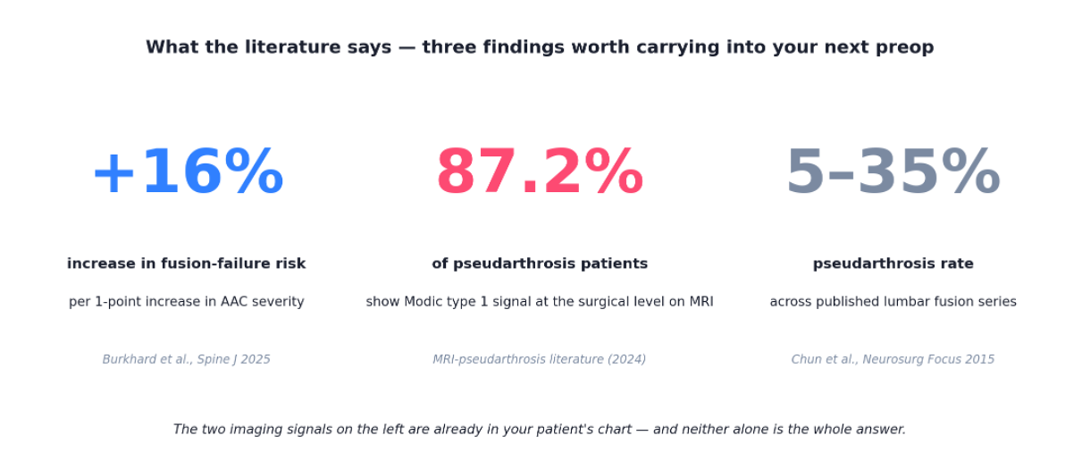
Pseudarthrosis remains one of the most common and costly complications of lumbar fusion. Across published series, nonunion rates range from five to thirty-five percent depending on technique and the number of levels [1]. The established risk factors — smoking, age, number of levels fused, diabetes — are well documented and routinely incorporated into preoperative planning, yet they do not fully account for the observed variance in surgical outcomes.
The unexplained variance has a measurable component, and an accumulating body of literature suggests that much of it is visible on routinely acquired preoperative imaging. Pseudarthrosis is not a stochastic complication. It is a calculation — and the majority of its inputs are already present in the patient record.
What the Established Risk Factors Do Not Explain
A 2024 systematic review and meta-analysis of lumbar fusion outcomes identified the dominant clinical and surgical predictors of pseudarthrosis. Smoking carries a relative risk of 1.91 compared to non-smokers, age is independently significant, and the number of fused levels is the dominant surgical predictor [2]. These findings, while well established, leave a substantial portion of the inter-patient variance unexplained.
If known clinical risk factors fully accounted for pseudarthrosis, the seven-fold range in published fusion-failure rates would not exist. The remaining variance is at least partially attributable to imaging biomarkers that are visible on routine preoperative studies but rarely scored — most notably abdominal aortic calcification (AAC) and Modic endplate changes.
Abdominal Aortic Calcification as a Predictor of Fusion Failure
Abdominal aortic calcification is visible on every lateral lumbar radiograph and on every preoperative CT, yet it is almost never scored as part of surgical risk assessment. The published evidence indicates that it should be.
Burkhard and colleagues recently published a 207-patient cohort in The Spine Journal demonstrating that AAC severity is an independent predictor of impaired posterolateral fusion after elective lumbar fusion [3]. Each one-point increase in the AAC severity score corresponded to a 16 percent increase in fusion-failure risk, and 28.5 percent of patients in the cohort demonstrated impaired posterolateral fusion. A related analysis from the same group has shown that AAC severity also independently predicts perioperative blood loss in posterior lumbar fusion [4].
The mechanism is biologically coherent. Vascular calcification reflects systemic atherosclerotic disease, including the small segmental vessels supplying the lumbar vertebrae. Bone healing is dependent on adequate vascular supply. When the vasculature serving the fusion bed is compromised — as evidenced by calcific deposition on routine imaging — the biological substrate required for solid arthrodesis is compromised in parallel.
Modic Changes as a Predictor of Nonunion
Modic changes — the type 1 inflammatory pattern (low T1, high T2) and the type 2 fatty pattern (high T1, high T2) — were originally described as endplate findings associated with degenerative disc disease. The literature on their relationship to surgical outcomes has accumulated steadily over the past three decades.
Buttermann's early observations and subsequent confirmatory studies established that nonunion after lumbar fusion is predominantly associated with the persistence of type 1 Modic changes, whereas solid fusion is associated with type 2 changes. A more recent MRI-based diagnostic series reported that 87.2 percent of patients with confirmed pseudarthrosis demonstrate a Modic 1 signal at the surgical level [5]. The pathophysiological interpretation is consistent: type 1 signal reflects ongoing endplate edema and inflammation — the radiographic signature of bone that has not consolidated.
Modic patterns are documented on the majority of preoperative lumbar MRIs and are routinely noted in radiology reports. Their use as a predictor of postoperative outcome remains uncommon despite the supporting evidence.
Composing a Multi-Variable Risk Model
No single signal — AAC, Modic, smoking, vertebral bone quality (VBQ), paraspinal muscle quality, or glycemic state — predicts pseudarthrosis with high accuracy in isolation. The published evidence suggests that composing these signals into a calibrated, multi-variable model is the appropriate next step. This is the design principle underlying Surgeon Decision Intelligence: a tissue-typing layer that extracts signals already present in preoperative imaging and clinical history, integrates them per patient, and outputs a calibrated probability the surgeon can act on at the bedside.

A risk indicator and a durability index which includes pseudoarthrosis does not principally determine whether to offer fusion. It informs the three adjacent decisions: whether patient optimization is needed, whether to augment biology at the time of surgery, and whether to reconsider the construct entirely — longer levels, a different approach, or non-fusion.
Conclusion
Pseudarthrosis has long been treated as an inherent risk of fusion surgery. The accumulating evidence suggests that it is, in significant part, a predictable one. The relevant signals are present in routinely acquired preoperative imaging and clinical data. The pending task is not the discovery of new biomarkers, but the systematic integration of those already in use.
References
[1] Chun DS, Baker KC, Hsu WK. Lumbar pseudarthrosis: a review of current diagnosis and treatment. Neurosurgical Focus. 2015;39(4):E10. PMID 26424334.
[2] Pseudarthrosis risk factors in lumbar fusion: a systematic review and meta-analysis. BMC Musculoskeletal Disorders. 2024. PMC11149252.
[3] Burkhard MD, et al. Abdominal aortic calcification is associated with impaired fusion after elective spinal fusion. The Spine Journal. 2025. PMID 40216361.
[4] Abdominal aortic calcification is an independent predictor of perioperative blood loss in posterior spinal fusion surgery. European Spine Journal. 2024. DOI 10.1007/s00586-024-08184-y.
[5] Contribution of MRI and imaging exams in the diagnosis of lumbar pseudarthrosis. ScienceDirect. 2024. (87.2% Modic-1-signal prevalence in confirmed pseudarthrosis). See also: Buttermann GR, et al. Vertebral body MRI related to lumbar fusion results. European Spine Journal. 1997. For background on Modic-fusion relationships.