
I've been thinking a lot lately about how we measure success in spine surgery. It's fascinating—and honestly a bit frustrating—how the goalposts keep moving. Just when we think we've figured out the perfect angle or the ideal measurement to predict good outcomes, someone publishes a new study showing us we've been missing something important.
Let me show you what I mean.
The Moving Target of Spine Surgery

It feels like every week there's a new paper introducing another angle, another measurement, another "key parameter" that we absolutely must consider for good spine outcomes. The goal posts keep moving.What was considered optimal alignment five years ago might be outdated today.What's considered cutting-edge today might be replaced by something else tomorrow.
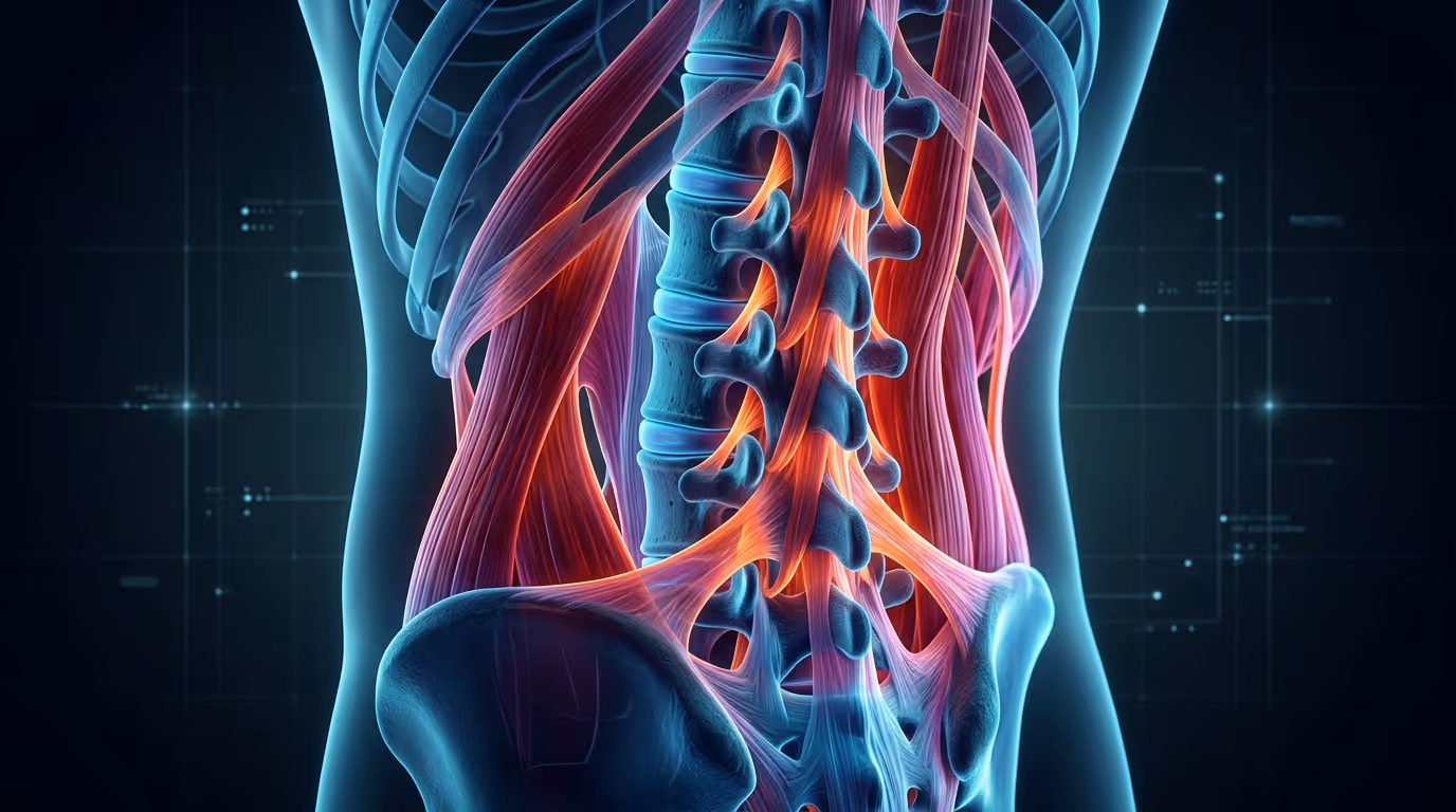
And here's what strikes me most about this continuous evolution: every single one of these metrics focuses exclusively on bones.We're measuring angles between vertebrae, distances between bony landmarks, the position of the pelvis relative to the spine. It's all bone.
And that's the problem.
Our current understanding of spine stability is built on an old architectural model—one that doesn't actually reflect how the spine works in real life. I want to explain this using a metaphor that completely changed how I think about spinal surgery.
Pyramids vs. Buildings: Rethinking Spine Architecture

For the longest time, we've been treating the spine like a pyramid. Think about the Egyptian pyramids—massive stone structures with wide, rigid bases. They're stable because of their geometry and the sheer weight of the stones. They don't bend. They don't adapt. They just sit there, immobile and heavy, relying entirely on static stability from the stone blocks stacked on top of each other.
That's the old paradigm. We looked at the spine as a stack of bones that needed to be perfectly aligned, and as long as we got the angles right, everything would be fine. It was all about load-bearing through bone alone, with passive support at best.
But here's the reality: we don't build pyramids anymore. We build skyscrapers.
Think about a modern high-rise building swaying in the wind.It doesn't topple over because it has a narrow, flexible core with dynamic support systems. The building can adapt to stress. It redistributes loads. And critically, it relies on much more than just its central concrete structure.
This is the new paradigm for understanding the spine. The spine isn't a rigid pyramid—it's a flexible tower that depends on dynamic soft tissue architecture:
- Muscles are the guy wires: Just like the cables that stabilize a tall antenna or suspension bridge, our paraspinal muscles actively pull and adjust to keep us upright. They're not passive—they're constantly working, responding to every movement we make.
- Ligaments are the internal trusses: Inside a skyscraper, you have steel framework and cross-bracing that distributes forces throughout the structure. That's what ligaments and fascia do. They connect vertebrae, limit excessive motion, and help transfer loads across multiple levels.
- Vascular and connective health are the foundational piles: Before you build a skyscraper, you drive deep foundation piles into the ground. These aren't visible, but without them, the whole building sinks. Similarly, the health of our blood vessels, the quality of our connective tissue, and the overall biological environment determines whether healing happens and whether implants stay secure.
When you think about it this way, it becomes obvious: you can't just focus on the bones. A building engineer wouldn't design a skyscraper by only thinking about the concrete core and ignoring the steel framework, the guy wires, and the foundation. That would be absurd. Yet that's essentially what we've been doing in spine surgery—focusing almost entirely on bony alignment while largely ignoring the soft tissue support system.
Why Have We Ignored Soft Tissue for So Long?
If soft tissue is so important, why hasn't it been a bigger part of the conversation until now? The answer is frustratingly simple: it's really hard to measure.
I can look at an X-ray and measure the Cobb Angle in about thirty seconds. I can pull up a sagittal X-ray and measure the SagittalVertical Axis just as quickly. These measurements are objective, reproducible, and easy to communicate. That's why classifications based on bony alignment are so popular in medical literature—they're simple, even if they're not always as prognostic as we'd like them to be.
But soft tissue? That's a completely different challenge.How do you measure muscle mass objectively? How do you quantify muscle quality versus just muscle size? What about fat infiltration into the muscles—how much is too much? And vascular calcification—how do you measure that consistently across different MRI platforms and different hospitals?
Even if you figure out how to measure these things, you're immediately drowning in information. An MRI scan contains millions of datapoints. Which ones matter? Does the muscle at L3 matter more than L4? Is the psoas muscle more important than the multifidus? What about the fat in the subcutaneous tissue versus the fat infiltrating the muscle itself? To complicate things further, differences between MRI machines makes it hard to compare images from one MRI to another.
Without a systematic way to analyze all this information, without artificial intelligence and big data approaches, it's nearly impossible for a single surgeon to comprehend and use this information in a meaningful way. So we've defaulted to what's easy: bones.
The Comprehensive Approach: What the Science Is Telling Us

Here's where things get really interesting. Recent research is showing us—in no uncertain terms—that soft tissue matters. A lot. Let me share some findings that I find absolutely compelling:
Low paraspinal muscle mass strongly predicts implant failure, with an AUROC (that's a statistical measure of predictive accuracy) of0.83. Think about that—the muscles around the spine predict hardware failure better than most of our bone-based metrics. In addition, low muscle mass correlates directly with inferior patient-reported outcomes.
Patients who develop adjacent segment disease have 24% more fat in their paraspinal muscles at L3. Adjacent segment disease is when the level above or below a fusion starts breaking down. We've always wondered why some patients get this and others don't. Turns out, fat infiltration in the muscles is a major clue. Those muscles aren't working properly, and the spine compensates by overloading the next level.
Aortic calcification independently predicts lumbar fusion failure. This is about vascular health—those foundation piles I mentioned earlier. If the blood vessels are calcified, healing doesn't happen properly.The bone doesn't fuse. The hardware loosens. It doesn't matter how perfect your alignment is if the biological environment can't support healing.
Joint effusion and disc degeneration predict treatment failure following decompression. Even in less invasive surgeries, the quality of the surrounding tissues matters. Inflammation in the joints, degeneration in the discs—these aren't just incidental findings. They're predictive of outcomes.
All of these studies point to the same conclusion: bone-only planning misses critical risk signals. If we want to get to a true gold standard in spine surgery—where we can reliably predict who will do well and who won't, where we can optimize treatment selection for each individual patient—we must think comprehensively. We need both the bones and the soft tissue.
The New Frontier: Big Data and Automated Analysis
So here's the exciting part: we're finally at a point where we can actually do this. Big data and artificial intelligence are making it possible to automatically analyze soft tissue from standard MRI scans. We can now quantify muscle mass, measure fat infiltration, assess vascular calcification, and evaluate dozens of other parameters in minutes—things that would have taken hours or been impossible just a few years ago.
By putting data online, sharing imaging databases, and developing machine learning algorithms, we can train computers to identify patterns that predict outcomes.
I believe we're on the cusp of a paradigm shift in spine surgery. In this new standard, soft tissue guides surgical treatment decision.I can look at someone's MRI and see not just their disc herniation, but also their muscle quality, their vascular health, their joint inflammation. All of those factors tell me something about their risk. Some patients are high-risk based on their soft tissue profile, and knowing that upfront changes the conversation.
Most importantly, soft tissue assessment can help produce longevity in spine health. If we understand that muscles, ligaments, and vascular health are critical to spinal stability, then we start thinking about preserving and optimizing those tissues. We think about physical therapy differently. We think about nutrition and metabolic health as part of spine care. We think about the spine as a dynamic, living system—not just a stack of bones.
The Big Takeaway
For decades, we've been chasing the perfect angle, the ideal measurement, the magical number that would tell us exactly how to fix someone's spine. And we've made progress—real progress. But we've been building pyramids when we should have been building skyscrapers.
The future of spine surgery isn't about finding yet another angle to measure. It's about comprehensive analysis that incorporates everything: bones, muscles, ligaments, vascular health, connective tissue quality.
The goal posts have moved many times over the past 70 years.But I think this time, we're not just moving them—we're building a new field entirely. And I'm excited to see where it takes us.